
Keratoconus Detection Center
What is Keratoconus?
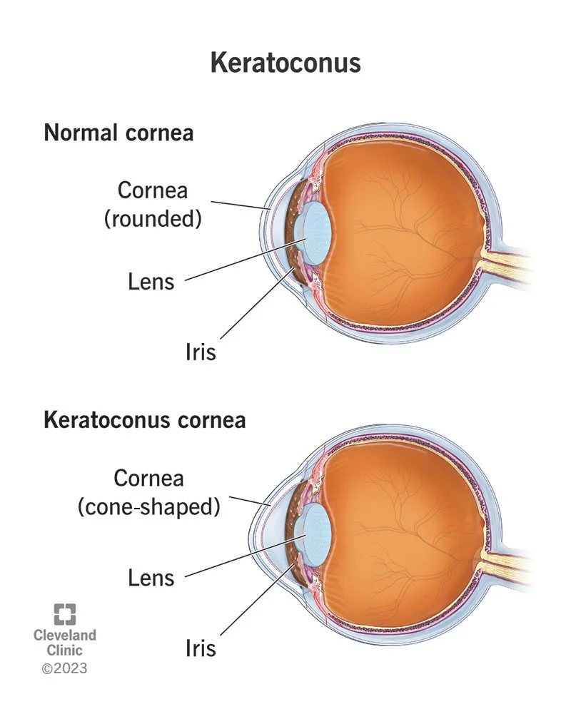
Keratoconus is an eye condition in which the normally round cornea gradually becomes thinner and bulges outward into a cone-like shape. The cornea is the clear front surface of the eye that plays a critical role in focusing light for clear vision.
Keratoconus is most commonly diagnosed in the teenage years or early adulthood, though it can begin in childhood or, in milder cases, be detected later in life. The condition typically progresses over time, with changes often occurring more rapidly in younger individuals.
As keratoconus advances and the cornea becomes more irregular, vision may become increasingly distorted. In many cases, specialty contact lenses—such as rigid gas permeable (RGP), scleral, or hybrid lenses—can provide clear, stable vision by creating a smooth optical surface over the cornea.
Prevention and Treatment?
Corneal cross-linking is a minimally invasive procedure designed to strengthen the cornea and slow or halt the progression of conditions such as keratoconus. Early treatment is key—cross-linking is most effective when performed soon after diagnosis, before significant changes in vision occur.
With newest FDA-approved advancements - the Epioxa corneal cross-linking procedure = can now be performed at the time of diagnosis rather than waiting for documented progression. This allows for earlier intervention and better long-term stability of the cornea.
At Princeton Optometry, we utilize advanced diagnostic technology to detect even subtle corneal changes. This is especially important for patients with a family history of corneal disease, as early screening can lead to timely treatment and improved outcomes.
Corneal cross-linking may also be recommended later in the course of the disease if there are signs of continued progression, such as increasing astigmatism or changes in glasses prescription.